This topic is about how different combinations of genes can express different features, and how these features can be inherited. It is a very important topic since it builds the basis for a lot of future chapters. I would therefore make sure to be as familiar with it as possible. The material itself is not all that difficult, but some of the problem-solving exercises can be. Practicing solving Punnett squares and pedigree charts is key to this chapter. The most challenging part about Punnett squares is to know what genotypes (representation of alleles with two letters) should be crossed. I will include a list of terms to make problem solving easier. Otherwise, remembering a few examples of different types of genetic diseases is probably a good idea.
Points for revision:
- List of terms for problem solving
- Human karyogram and chromosomes
- Punnett squares and inheritance
List of terms for problem solving
Heterozygous: Two different alleles, for example Bb
Homozygous: Two of the same alleles, for example BB
Recessive allele: Will not be expressed unless it is homozygous recessive, denoted by lower-case letter
Dominant allele: Will be expressed as long as it is present, denoted as upper-case letter
Genotype: Combination of alleles, denoted by two letters, for example Bb or BB
Phenotype: The trait expressed, for example round pea or wrinkled pea
Autosomal: genes not located on a sex-chromosome (chromosome pair 1-22)
Sex-chromosome: Chromosome pair 23, either XX (female) or XY (male)
Gamete: Sex cell, haploid
Carrier: A person carrying an allele for a disease, but is not affected
Genes
A gene is a distinct hereditable sequence of DNA coding for a specific characteristic. DNA is in itself a sequence of bases coding for amino acids, which codes for proteins that goes together and makes up what we are. Genes often have different versions of itself, and these versions are called alleles. The color of peas for example, is determined by a gene that either codes for green peas or yellow peas. The genes coding for green and yellow are called alleles of that gene. Furthermore, when the cell prepares for mitosis and supercoils its DNA into chromosomes, genes can be identified at the same location on the same chromosome every time. The location where a gene resides is called the loci of that gene.
Mutations
Genes are very specific, and a change in just one nucleotide in the DNA sequence, can completely alter the appearance of the characteristic the gene codes for. Changes in the DNA sequence are called mutations, and it can happen in various ways. A base substitution mutation for example is when a single nucleotide is replaced by another nucleotide. This is extremely rare since it is energetically unpreferred, but when it does happen, it might have major consequences. Sickle cell disease is an example of a base substitution mutation. Sickle cell disease occurs when an adenine is replaced by a thymine, which completely changes the structure of the protein it codes for. In the case of sickle cell disease, the proteins are red blood cells, and the change in structure causes the cell to fold in on itself, making a sickle shape. The consequences of the change are among other things that the sickle shaped cells easily tangle up with each other, blocking blood vessels and causing pain and swelling. However, it turns out that sickle cell disease also has its benefits, because the parasite which causes malaria cannot use sickle cells as its host. This gives people affected by sickle cell disease an immunity against malaria, which explains the high ratio of sickle cell disease in counties affected by malaria such as African countries. Mutations can therefore be both a benefit and a drawback.
2.jpg)
2.jpg)
Karyograms and chromosomes
A karyogram is a representation of the chromosomes of a species, ranged by size and shape. A chromosome is tightly wound DNA and is formed before cell division. DNA is first coiled around 8 protein structures called histones. The coiled DNA is called chromatin, and it is the form DNA takes when the cell is not dividing. Chromatin looks like a long chain of beads, and each bead is called a nucleosome. An additional histone is often present to make supercoiling possible. When the chromatin is supercoiled, it takes the form of a chromosome.
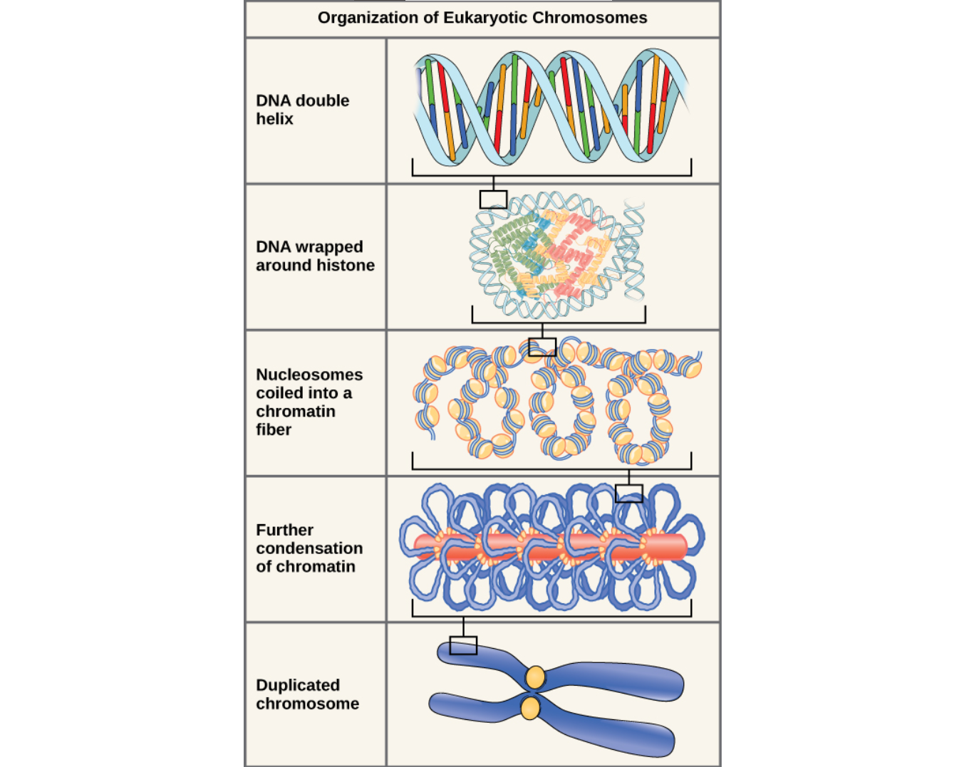
A diploid human cell has 46 chromosomes formed into 23 pairs. Half of the chromosomes are from the mother, and the other half from the father. Diploid cells are said to be 2n, since the number of chromosomes is doubled relative to haploid cells. Haploid cells in humans are referred to as sperm or egg cells. These cells divide by meiosis and thus only have half the chromosomes of a diploid cell. It is therefore said to have n chromosomes. Chromosome pair number 23 in humans are the sex determining chromosomes. If chromosome pair number 23 is XX, it is a female, and if it is XY, it is a male. Anomalies present in the karyogram, for example a different number of chromosomes, may result in genetic diseases. The most known genetic disease caused by a different number of chromosomes is downs syndrome. Downs syndrome is caused by errors in meiosis I or II.
Punnett squares and inheritance
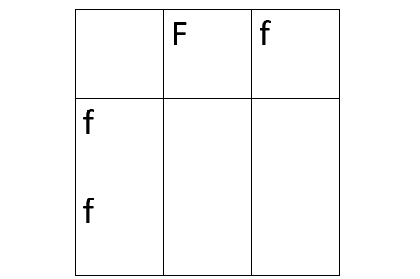
A Punnett square is a tool that can be used to determine the possible outcomes of a cross, and the probabilities of each outcome. To set up a Punnett square, the possible alleles carried by the parent gametes has to be determined. If a person has an allele F from the mother, and an allele f from the father, the genotype of that person is said to be Ff. Since dominant genes are denoted as capital letters, the person would express the phenotype of F. If this person would have a child, either f or F can be passed on due to meiosis and production of gametes. Let’s suppose that this person is having a child with someone with a genotype of ff. The possible gametes would then be f or f, and the Punnett square setup would look like this:
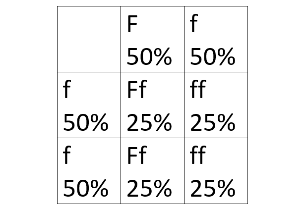
Since each gamete has a 50% probability of being the gamete inherited, each square in the cross has a 25% probability of being the genotype of the outcome. From the resulting Punnett square we can deduce that 50% of the outcome will express the phenotype of f, and 50% will express the phenotype of F.
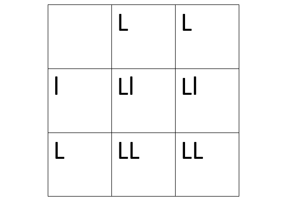
Suppose we are plant breeders and want to know whether a plant is purebred for a specific trait. We can then use test crossing to determine the genotype of the plant. Let’s say that we are looking for plants with wide leaves. We have deduced experimentally that wide leaves is a dominant trait, so we will denote is as L. We can then breed the plant with any other plant known to produce both wide and narrow leaves. If the outcome is 100% wide leaves, it can be deduced that the plant is true breeding for wide leaves, in this case homozygous dominant.
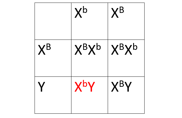
Some alleles are also sex linked, which means that the genes coding for the allele lies on a sex chromosome. Men are most affected by sex linked diseases because men have both a X and a Y chromosome, so any disease present on either chromosome is expressed. Women have two X chromosomes, so one allele can dominate the other allele if the disease is recessive. The gene for baldness is a recessive allele that lies on the X chromosome, which means that men who inherits a X chromosome with baldness is guaranteed to express the phenotype. Sex linked traits are denoted as the chromosome it is present on, and the symbol for the trait as a superscript. If we call the allele for baldness b, the way to represent the allele would be Xb.
Meiosis
Meiosis happens in a similar fashion as mitosis, but with a few distinct differences. Meiosis is called reduction division because unlike mitosis which produces 2 cells with 2n chromosomes (diploid), meiosis produces 4 cells with n chromosomes (haploid). Meiosis occurs in two main stages with 4 substages. The 4 substages has the same names as in mitosis: Prophase, metaphase, anaphase, and telophase. However, in meiosis it is called prophase 1, metaphase 1, … and then prophase 2, metaphase 2 … Both interphase and cytokinesis happen the same way for both mitosis and meiosis. Meiosis 2 happens in a fashion similar to mitosis, but meiosis 1 is special and has some important steps that does not happen in mitosis.
During prophase 1 of meiosis 1, the chromatin condenses to chromosomes, and then the chromosomes undergo recombination, also called crossing over. The two homologous chromosomes exchange genetic material, thus increasing genetic variation. The chromosomes then line up at the equator during metaphase 1 and the nuclear envelope disintegrates. However, the chromosomes do not line up in one straight line as in mitosis, instead they line up in two lines of homologous chromosomes (chromosome 1 with chromosome 1 and so on). Additionally, the chromosomes are not lined up in any specific order, it is completely random which of the homologous chromosome lies in which side of the division. This is called random orientation, which further increases genetic variation. Random orientation means that it is random if one specific chromosome is from the mother or the father. Anaphase happens the same way as mitosis, but the centromere is not split, meaning that the sister chromatids are still connected. They are not split until anaphase 2. Telophase 1 and cytokinesis happens the exact same way as mitosis. If the stages of mitosis are not clear, try to refer back to topic 1.
After meiosis 1, two cells with n chromosomes are formed, with crossing over and random orientation increasing the genetic variation of each cell. This means that the probability of two cells being identical is very small. The cell is now ready for meiosis 2. During meiosis 2, the two haploid cells still with its two sister chromatids attached, separate into 2 again, forming 4 cells with one chromatid each. Since the sister chromatids were not separated in meiosis 1, there is no need for another S phase between meiosis 1 and 2. Meiosis 2 happens in basically the same way as mitosis. In prophase 2, the chromosomes are condensed, in metaphase 2, the nuclear envelope disintegrates, and the chromosomes are lined up with random orientation. In anaphase 2, the chromosomes start to separate as the microtubules shorten. And the sister chromatids are separated as the centromere disintegrates. Finally, in telophase 2 the chromosomes are in opposite poles, and the cell elongates in preparation for cytokinesis. In cytokinesis the cell membrane in divided into two parts, and the final result is 4 haploid cells, where crossing over and random orientation has made sure that no two cells are identical.
Gametes (sex cells) do not undergo the cell cycle again. After meiosis 2, they are either paired up with another sex cell, or they undergo cell death.

Gel electrophoresis
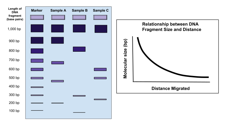
Gel electrophoresis is a technology which makes identifying DNA easier. DNA is cut into segments by enzymes and passed through a gel with a current. The smallest and most charged particles will travel the farthest, and the biggest least charged particles will travel the shortest. The resulting pattern can be compared to a known pattern to see if there is a match. Gel electrophoresis is often used in DNA profiling to compare a known DNA segment with suspected matches. If the two resulting patterns are identical, it is a match. It is useful in identifying suspects from a crime scene, or to find out who the biological parents of a child is.
Helpful videos:
Punnett squares: https://www.youtube.com/watch?v=agQpPPQ5IVQ
Pedigree charts: https://www.youtube.com/watch?v=bL4JV5Z2jsc
Meiosis: https://www.youtube.com/watch?v=ijLc52LmFQg
Human genome project: https://www.youtube.com/watch?v=MvuYATh7Y74
I experienced this chapter as informational and stuffed with real world applications of genetic technology, and the information was comprehensive and structured. However, Norwegian education on genetics is apparently quite good, since I knew most of the information beforehand. I therefore may not have found it as surprising as someone who might learn about this for the first time.